
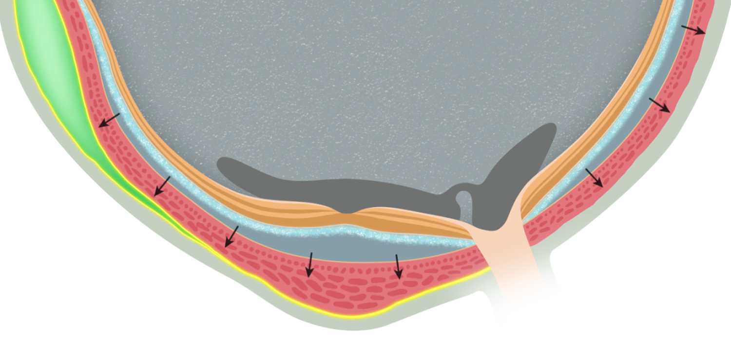
Dr. Rajeev Muni (vitreoretinal surgeon at St. Michael's Hospital, The Hospital for Sick Children and the Kensington Eye Institute) and his research team asked me to help them visualize a novel, minimally invasive procedure for a specific type of retinal reattachment.
Dr. Muni’s team has investigated how retinal detachment causes morphological changes in the retina. The growing literature around retinal pathomorphology has helped surgeons create more finely-tuned surgical plans and benchmarks for success. The goal for this visualization was to show a specific novel reattachment procedure within the context of 1) overall retinal reattachment, and 2) tissue-specific recovery.
This video accompanied the article Suprachoroidal Viscopexy for Chronic Rhegmatogenous Retinal Detachment Repair with Inferior Breaks, published in the Canadian Journal of Ophthalmology (Volume 60 Issue 5).
Suprachoroidal viscopexy for retinal detachment repair
-
Scholarly community working on retinal pathomorphology and retinal detachment procedures.
-
Storyboarding, illustration, animating, and compositing.
-
Dr. Rajeev Muni, Dr. Aurora Pecaku. Dr. Isabela Martins Melo
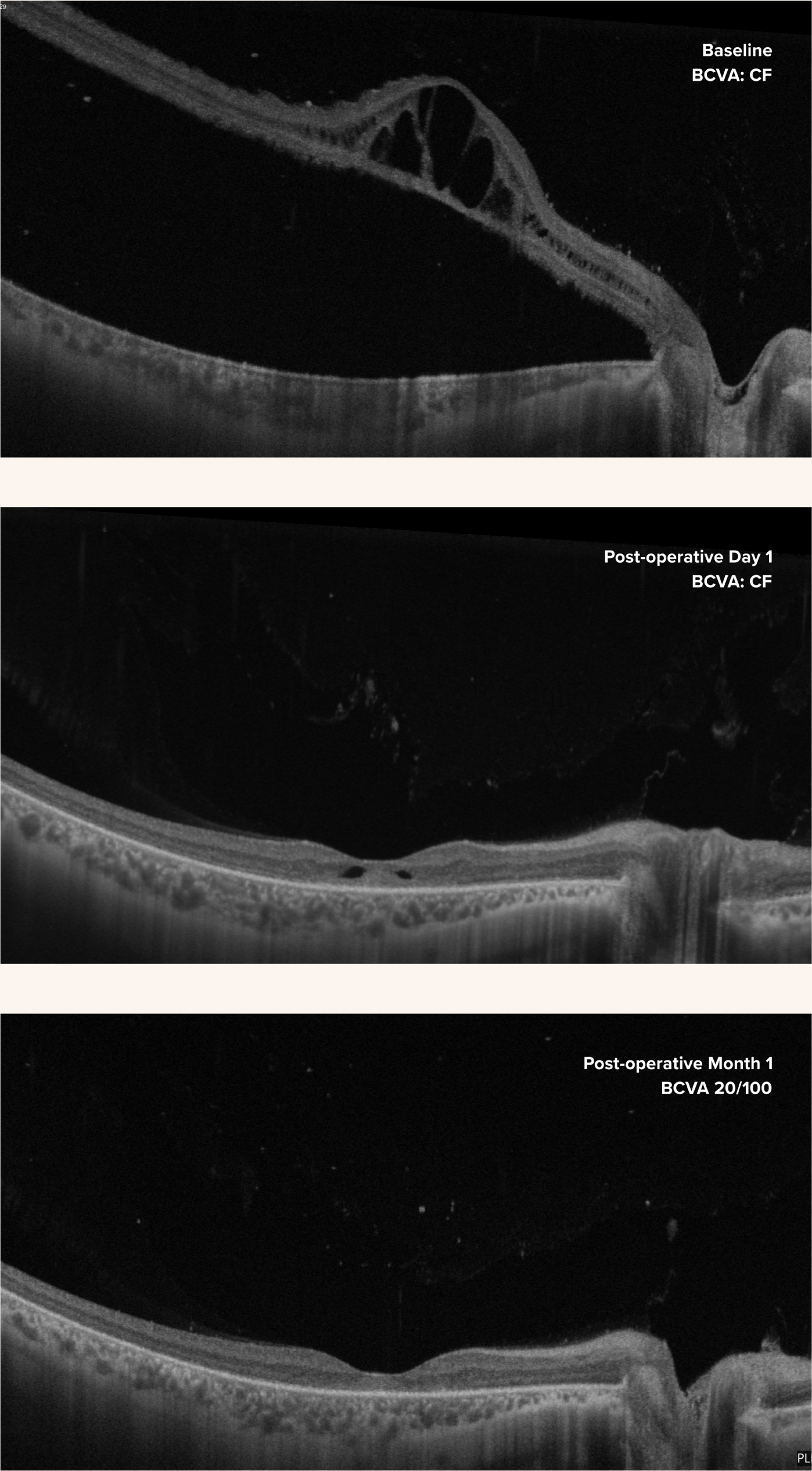
Optical Coherence Tomography (OCT) is a non-invasive imaging technique which produces highly detailed cross sections of the retina. OCT scans are a critical tool in the vitreoretinal surgeon’s toolkit, particularly when creating a surgical plan based on retina pathomorphology.
However, it is hard for OCT scans to single-handedly communicate tangible research insights. For one thing, every patient case is unique. Dr. Muni’s team has analyzed several individual patient cases, in order to parse clinically relevant commonalities from unique case-specific features. They have successfully determined a 5-part staging series to describe the progression of retinal detachment.
Second, while OCT scans are fairly straightforward to read, it can still be hard to parse what is happening in small areas of tissue (especially when the tissue is damaged).
Finally, OCT scans are specific snapshots in time (baseline, 1 day post-operation, etc). This can make it difficult to picture and compare progressive changes in the tissue across time, especially when these changes are happening on several levels of detail.
Our main approach to creating this visualization was to bridge the gaps between OCT scans. We recreated an OCT-style view of the retina so that it:
Was standardized and visually salient
Clarified clinically important visual markers and stages, as determined by a team of expert researchers
Showed an animated progression of the retina from detached to reattached
This animation drew on much of the team’s incredible work observing and categorizing stages of retinal detachment. It provides a view that cannot be afforded by OCT scans alone, as it clarifies the visual findings and links them across time.
Bridging a Surgical and Diagnostic View
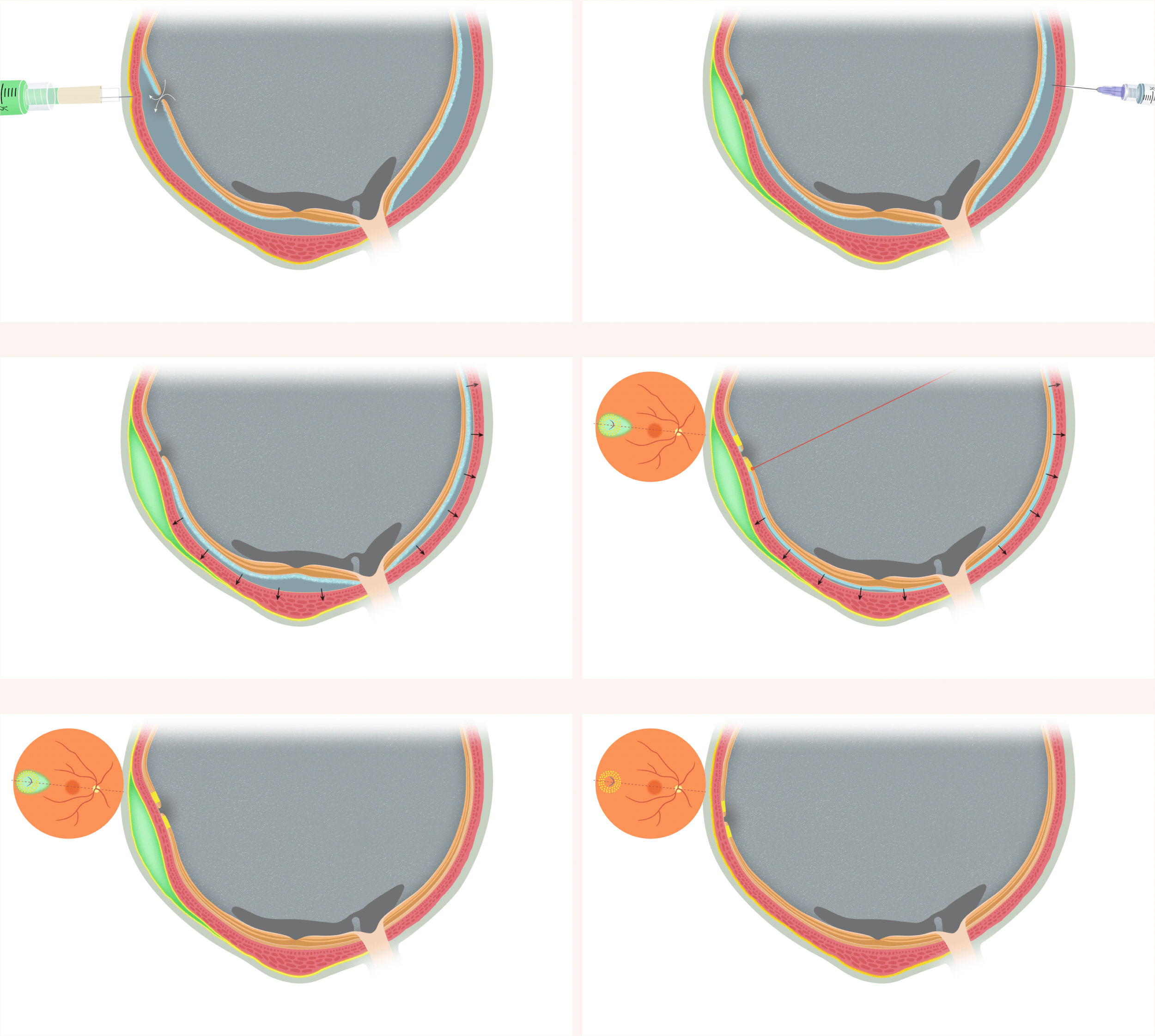
Translating Medical Images and Expertise into Motion
An illustrated OCT-style view of the retina was created based on the specifics of the case featured in the journal article (chronic/regulated rhegmatogenous retinal detachment with inferior retinal breaks). The illustrated OCT view was designed to balance naturalism with clarity. Natural tones of pink, brown and cream were used for the retinal layers, except for the ellipsoid zone (EZ), the retinal layer closest to the fluid as it seeps in under a retinal tear. When this happens, the EZ gets hydrated by the fluid. In the illustrated version, during its hydration the EZ turns blue. This helps to underscore the underlying process (blue = hydration) and improve visibility of this layer, which is where most of the dramatic morphological changes occur.
In an OCT scan, layers of the retina are captured in greyscale. However, some noncontiguous tissue layers appear as the same value on an OCT scan. We wanted viewers of our visualization, who would most likely be familiar with OCT imaging, to be able to understand our visualization with as little friction as possible. Therefore, any layers which appear as similar tones in an OCT were made the same colour in our visualization.
We worked on clearly showing the morphological changes in the retinal layers during reattachment, and synchronizing this with the broader stages of the surgical procedure. The animation had to be kept as editable as possible, as the decisions around how to clarify and present these morphological stages continued to evolve.
The tool used for injecting viscoelastic into the suprachoroidal space was created by Dr. Muni. Visualizing this tool involved synthesizing photos, recordings, descriptions and measurements. The procedure itself was visualized based off of descriptions and close review sessions with the team.
We ended up with a visualization that interfaces smoothly with medical images commonly used by retinal surgeons. This animation forms a bridge between a novel surgical procedure and OCT-derived insights into retinal pathomorphology.